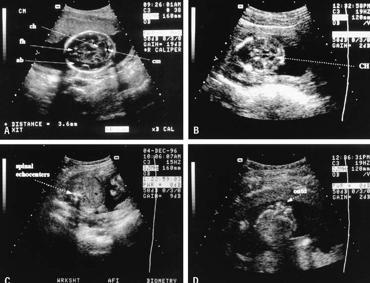
An ultrasound test is a radiology strategy that utilizes high-recurrence sound waves to create pictures of the human body’s organs and constructions. Ultrasound scanners comprise a PC, a presentation screen, and a transducer test to check the body. The transducer is a little hand-held gadget appended to the scanner. It is straightforwardly positioned on top of the territory to be observed, on which a greasing up gel is to be applied. Inward constructions at that point replicate the transducer’s sound waves through the body as “echoes.” These echoes get back to the transducer showing ongoing visual pictures on the presentation screen sent electronically.
Ultrasound pictures depend on similar speculations engaged with the sonar utilized by bats, ships adrift, and anglers with fish indicators. As a controlled sound bobs against objects, its repeating waves can be used to see its distance, shape, size, and internal consistency. A coordinated flood of quieted, high-recurrence sound waves into the body when the transducer is squeezed against the skin. The transducer records little changes in the sound’s pitch and heading when the sound waves reverberate from the body’s liquids and tissues. These waves are estimated and showed by a PC, which produces an ongoing picture on the presentation screen.
Obstetric ultrasound filtering is an ultrasound imaging strategy intended to expand actual assessments throughout pre-birth care. It has gotten very normal for guardians to demand print-outs of the pictures of their developing newborn child. The specialist now and again prints out images to see and clarifies the hatchling’s design as seen on screen to the guardians during the obstetric ultrasound check.
In ultrasound imaging, high-recurrence sound waves are ricocheted off the body to make a precise picture of the uterus. This is accomplished using a transducer that radiates waves and produces an image dependent on the reaction time’s length and recurrence adjustments. The ultrasound results made can be either a still or moving picture, with trendsetting innovation being actualized to make three-dimensional ultrasound pictures that give significantly more explicit subtleties. The obstetric ultrasound picture might be procured by covering the lady’s midsection in a conductive gel and running the transducer along the gut or embedding the transducer into the vaginal channel to get a clearer picture of a transvaginal ultrasound. The following picture gives an image of the uterus and its substance, alongside nearby body structures. These estimations diagram the establishment in evaluating gestational age, size, and development in the baby. A full bladder is regularly mandatory for the strategy when stomach filtering is done in the early pregnancy stages. There might be some uneasiness from tension on the full bladder.
There are a wide assortment of employments for it. Its imaging is generally used to assess pregnancy. This may incorporate deciding how far along the pregnancy is and affirming that the hatchling is regularly growing. Developments, for example, fetal heartbeat and irregularities in the hatchling, can be assessed, and estimations can be made precisely dependent on the screen’s pictures. An ultrasound can likewise be utilized explicitly to check for fetal abnormalities or issues, including a withdrawn placenta. Assume a mother accompanies pregnancy difficulties showing fetal misery. It could be used as an analytical instrument to beware of the infant’s status without utilizing intrusive strategies to imperil the pregnancy.
As there are various obstetric applications, specific obstetric ultrasound tests are required, contingent upon which is shown. On the off chance that an usb ultrasound scanner model has fixed missions, it might just be appropriate for a restricted subset of utilizations. Hence, it is regular for ultrasound frameworks to have tradable tests, and they habitually have more than one test association attachment for various applications.

